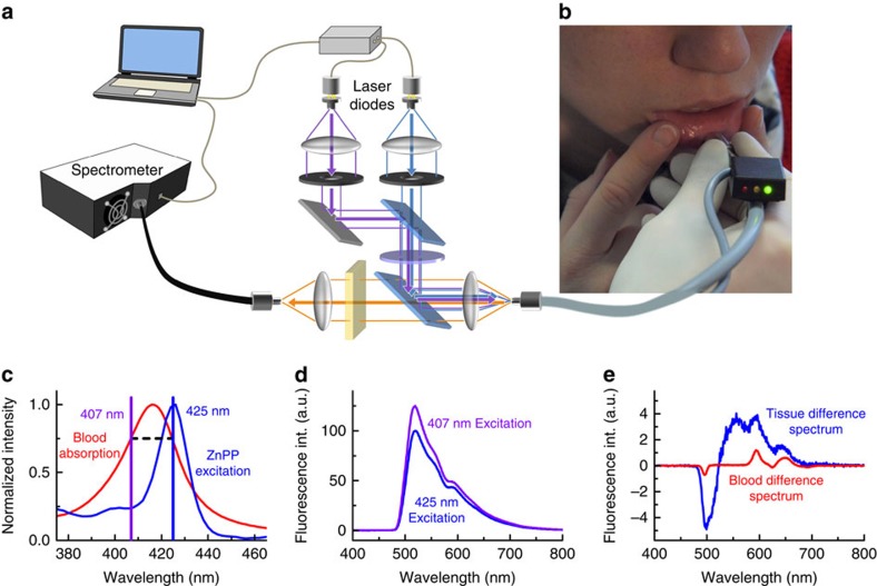
Figure 1. Tissue fluorescence measurements using dual-wavelength excitation.
(a) Simplified schematic of the set-up. Light of two laser diodes at 407 and 425 nm is superimposed and coupled into an optical fibre for fluorescence excitation. The generated fluorescence is collected by the same fibre and detected with a spectrometer. (b) Photograph of the measurement procedure with the fibre-optic probe placed on the lower lip. An adequate blood absorption index is indicated by the green light on the black indicator box. (c) The fluorescence excitation maximum of red blood cell zinc protoporphyrin (ZnPP) is at 425 nm. The second excitation wavelength of 407 nm is chosen for identical blood absorption. (d) Typical fluorescence spectra of the oral mucosa of a single subject with elevated ZnPP/haem ratio when excited with 425 and 407 nm. (e) Difference spectrum of the spectra in d with normalized 407-nm spectrum, resulting in background reduction of ∼93%. The corresponding difference spectrum of a measurement in whole blood using the same instrumentation in vitro is shown in red, obtained by placing the optical fibre in a sample of whole blood.

