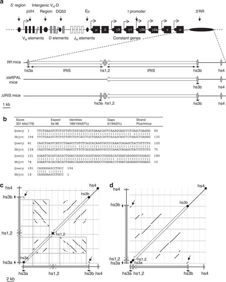
Figure 1. Palindromic structure of the IgH 3'RR.
(a) Upper part: The mouse IgH locus (not on the scale). Lower part: The 3'RR with its four enhancer elements and the IRIS (on the scale). ΔleftPAF and ΔIRIS mice are represented. (b) Sequence and homology between hs3a and hs3b enhancers (in opposite orientation in the chromosome). (c,d) DNA sequence dot-plot of the 3'RR in wt (c) and ΔIRIS mice (d) showing self similarity. The main diagonal represents the sequence alignment with itself. Parallel lines to the main diagonal represent repetitive patterns within the sequence (that is, tandem repeat), whereas perpendicular lines to the main diagonal represent similar but inverted sequences, thus allowing to identify the palindromic structures (dotted lines). The inversion of hs3a enhancer (black arrows) and the deletion of the intervening sequences between hs3a and hs1,2 in ΔIRIS mice totally disrupts the palindromic structure, despite the presence of all enhancer elements.

