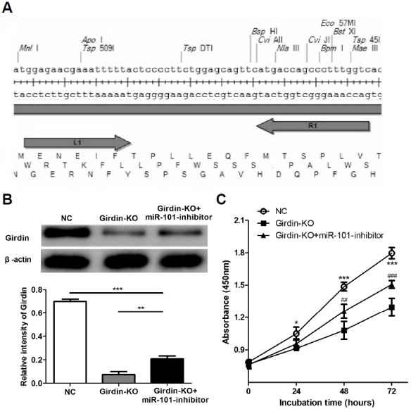
Fig. 5.

Silencing Girdin expression inhibited proliferation ability of HepG2 cells. (A) The plasmid structure of Talen vector. (B) HepG2 cells were transfected plasmids in order to knockout of the Girdin gene. Forty-eight hours later Girdin expression levels were determined by western blotting. (C) CCK method was used to detect proliferation of HepG2 cell at indicated time points. *P < 0.05, ***P < 0.001 (NC vs. Girdin-KO), ##P < 0.01, ###P < 0.001 (Girdin-KO+miR-101 inhibitor vs. Girdin-KO).
