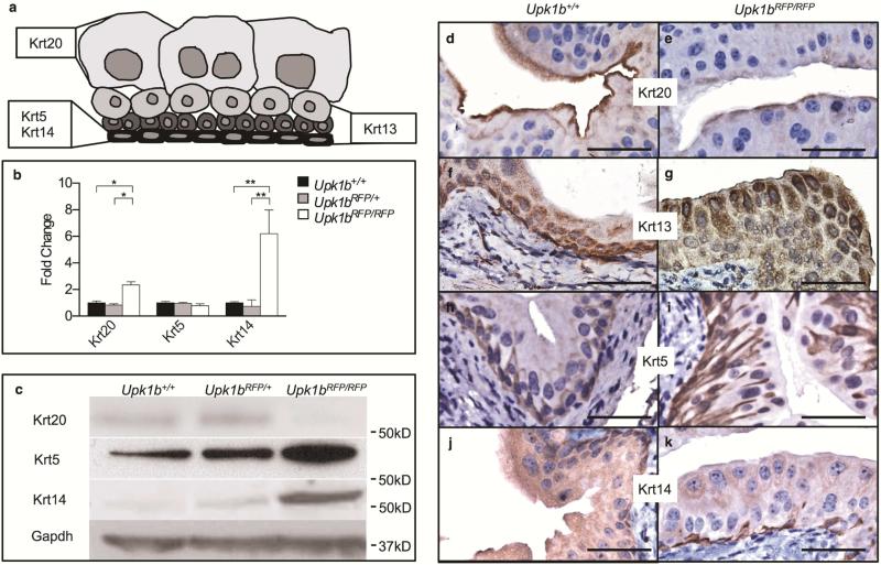
Figure 3. Absent Upk1b and Urothelial Plaque Affect Urothelial Homeostasis, Maturation and Organization.
(A) Schematic of urothelial cell morphology including the previously reported cytokeratin (Krt) profiles of basal, intermediate and umbrella cells. The cytokeratins used for immunohistochemistry are indicated in bold. (B) Cytokeratin mRNA expression in Upk1b+/+, Upk1bRFP/RFP and Upk1b+/+ bladders (n=5) was analyzed by qPCR. Raw data was normalized to Gapdh and fold change relative to Upk1b+/+ is graphed. A Two-Way ANOVA and Tukey's multiple comparison post-hoc test were used to evaluate statistical significance. *p < 0.05, **p < 0.01. Error bars represent standard error. (C) Protein expression in Upk1b+/+, Upk1bRFP/+ and Upk1bRFP/RFP bladders was evaluated by immunoblotting for the antibodies indicated. (D-K) Urothelial morphology in Upk1b+/+, Upk1bRFP/RFP bladders was analyzed by immunohistochemistry for the antibodies indicated. Scale bars indicate 25μm.

