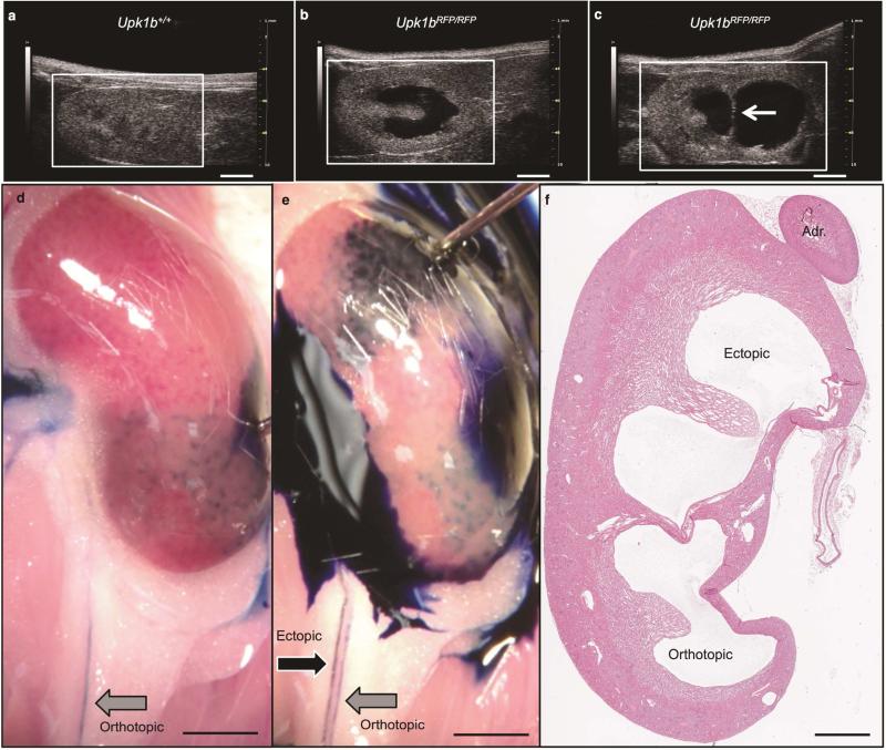
Figure 8. Identification of a Duplicated Collecting System.
(A-C) Renal ultrasound was used to evaluate Upk1b+/+ and Upk1bRFP/RFP adult mice to discern (A) normal renal structure, the presence of (B) hydronephrosis, or (C) hydronephrosis and collecting system duplication. White Arrow: Echogenic strip separating two renal pelvises. (D, E) Gross evaluation paired with methylene blue injection directly into the orthotopic (bottom, D) and ectopic (top, E) renal pelvises was used to identify two parallel ureters exiting the kidney. Black Arrow: Ectopic Ureter, Gray Arrow: Orthotopic Ureter. (F) The presence of ectopic and orthotopic pelvises, was analyzed by H&E staining. Adr (Adrenal Gland). Scale bars indicate 2mm, (A-E) 1mm (F).

