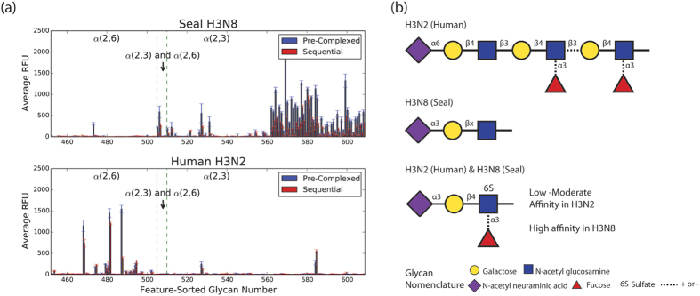
Figure 4. Glycan binding profiles of human H3N2 and seal H3N8 recombinant HA proteins.
(a) The glycans on our array were sorted according to the type of their sialic acid linkages (X-axis) and plotted against the averaged relative fluorescence unit (RFU) values (Y-axis). Error bars represent SEM of 4 RFU values for each glycan tested in our array. (b) Glycan cartoons representing the most prevalent motif bound by either H3N2 HA (top), H3N8 HA (middle) or both H3N2 and H3N8 HA (bottom). Dotted lines indicate mixed presence and absence among commonly bound glycan motifs.

