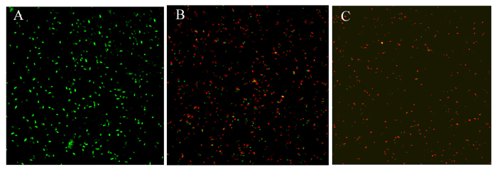
Figure 6. Confocal fluorescent microscopy images of live and dead Xoo cells after incubation with different samples.
Fluorescence image of Xoo treated with control (A); Fluorescence image of Xoo treated with Ag NPs (B); Fluorescence image of Xoo treated with SiO2-Ag composites (C). Green spots represent live bacterial cells, whereas red fluorescence indicates dead bacteria.

