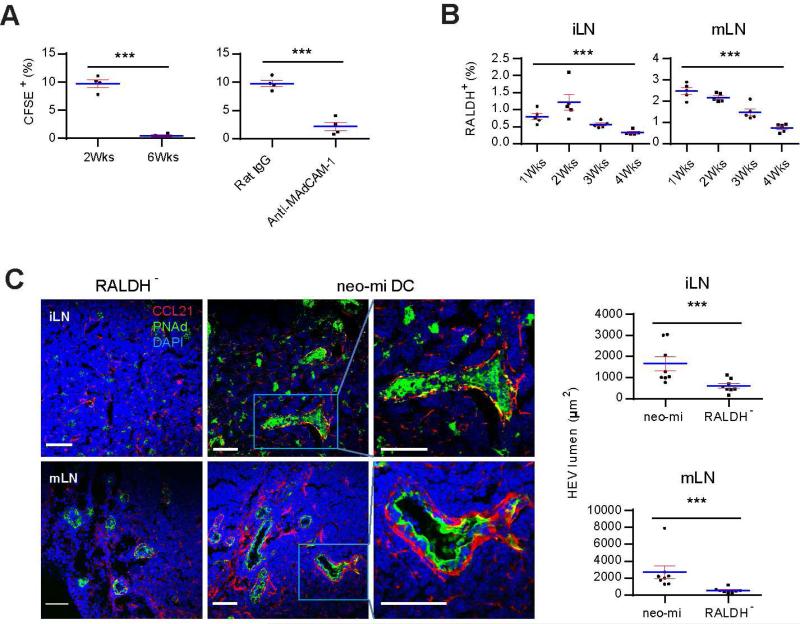
Figure 5. Neo-mi DCs induce neonatal to adult pLN addressin switch and HEV portal enlargement.
(A) Homing of neo-mi DCs into 2 and 6 week-old iLN. Left panel, 2 × 10⁵ CD45+RALDH+ neo-mi DCs were isolated from pLN of 2 week-old SPF mice and labeled with CFSE, and their migration to 2 and 6 week-old iLN was analyzed after 24 hrs as a percentage of total CD103+ cells in LN. Right panel, similar to the left, 2 week-old mice were injected with MAdCAM-1-blocking antibody as detailed in the methods, the migration of neo-mi DCs to iLN of 2 week-old mice after 24 hrs was compared to a rat IgG-injected control. The data are representative of three independent experiments. (B) The percentages of neo-mi DCs in iLN and mLN of 1, 2, 3 and 4 week-old SPF mice were analyzed by flow cytometry. The data are representative of at least five independent experiments. (C) 2 × 10⁵ neo-mi DCs were isolated from 2 week-old iLN of SPF mice, and i.v. injected into tail veins of 5 week-old GF mice in containment. The expression of PNAd and CCL21 in iLN and mLN was analyzed by confocal imaging after 7 days. Right panels: HEV portal areas (pixels surround by PNAd) were counted from at least 8 random images (3 mice per group). The images are representative of at least 30 photos. The statistics are shown from the pooled data. Also see Figure S5.

