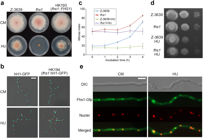
Figure 2. Fungal specific transcription factor Fhs1 involved in hydroxyurea sensitivity.
(a) Mycelia growth of F. graminearum strains on CM and CM supplemented with 10 mM HU. Pictures were taken 4 days after inoculation. (b) Nuclei and septa formation in hyphae. Conidia of both strains were inoculated into CM and CM supplemented with 10 mM HU for 24 h. Histone H1 was tagged with Gfp to visualise nuclei in both wild-type and fhs1 mutant strains. Septa confirmed by Calcofluor white staining are indicated with red bars. DIC and fluorescent protein images were merged. Scale bar = 20 μm. (c) Mitosis assay. Germlings bearing cells with two or more nuclei for 4-h incubation with and without 100 mM HU were scored. One hundred conidia were assessed for each strain with three biological replicates. (d) Serial dilutions of all strains were point-inoculated onto CM after 4-h incubation with or without HU treatment (106, 105, and 104 conidia/ml). (e) Subcellular localisation of Fhs1. Fhs1 was fused with Gfp, and histone H1 was fused with Rfp. Mycelia were harvested for microscopic observation 12 h after conidia inoculation in liquid CM supplemented with or without 5 mM HU. The yellow colour in the merged images indicates colocalisation. DIC, differential interference contrast. Scale bar = 10 μm.

