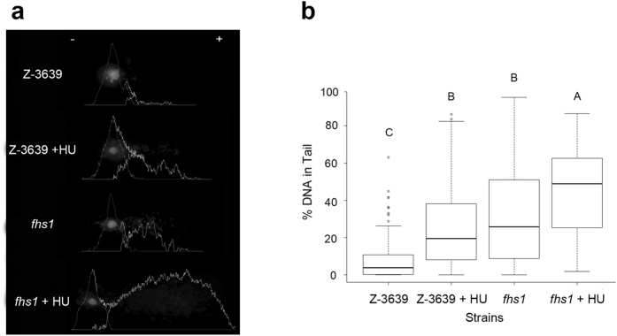
Figure 4. Detection of increased DNA damage.
(a) Photographs of comets and analysis of the alkaline comet assay. DNA fragments generated from DNA single-strand breaks, double-strand breaks, and alkali-labile sites in the DNA migrate toward the anode, creating the comet assay. Between the comets of fhs1 and HU-treated wild-type, fhs1 cells produced longer tails. Graphs depict image analysis quantifying the DNA contents of the head and tail. (b) Box-plot of average percentages of DNA in comet tails. fhs1 cells contained higher percentages of DNA in the comet tail than wild-type but similar to HU-treated wild-type cells (P < 0.01). HU-treated fhs1 cells contained significantly higher percentages of DNA in the comet tail than wild-type and HU-treated wild-type cells (P < 0.01).

