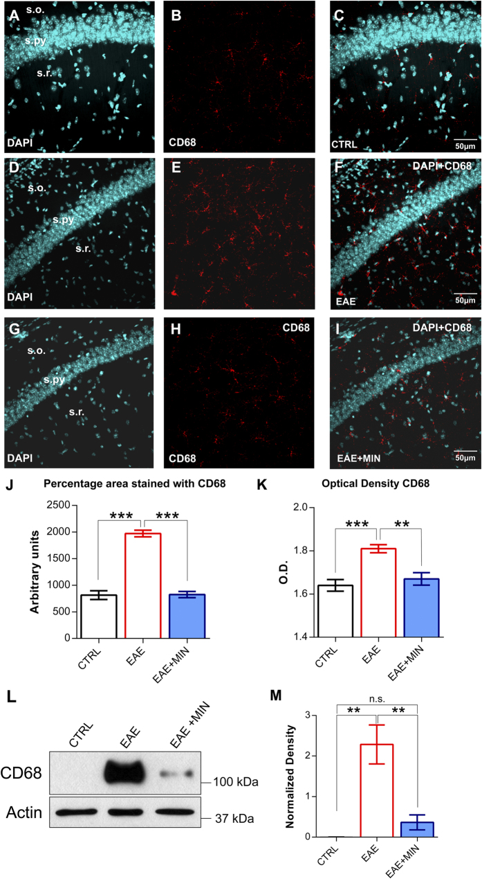
Figure 1. Hippocampal microglia activation in the remission phase of EAE is prevented by minocycline treatment.
(A–I) Confocal laser scanning microscopy (CLSM) images of double-label immunofluorescence for DAPI (visualized in blue) and CD68 (visualized in red-cy3 fluorescence) in the CA1 hippocampal region from control mice (A–C), EAE mice in the remission phase (D–F) and minocycline-treated remitting EAE mice (G–I). (J) Histogram showing the microglial reactivity quantification in terms of area immunolabeled by CD68 in control mice, remitting EAE mice and remitting EAE mice treated with minocycline. Data are presented as the mean values of CD68 positive areas ± SEM. Please note that the intense microglial reaction observed in the remission phase of EAE is prevented by the treatment with minocycline. (K) Histogram showing the microglial activation in terms of optical density for CD68. Each value is the mean ± SEM. (L) Western blot analysis of CD68 in the three experimental groups (n = 3 for each group). (M) Analysis of the actin-normalized optical density of the bands showed a significant increase of CD68 expression in EAE mice when compared to controls and subsequent decrease of the expression after minocycline treatment (mean + SEM). The reported P-values are relative to within group comparisons (Tukey’s range test, ***P < 0.001, **P < 0.01, *P < 0.05).

