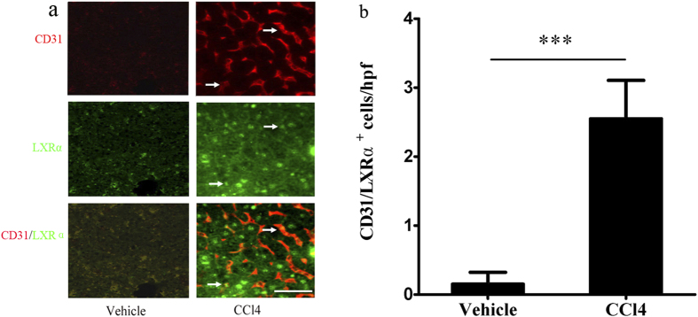
Figure 1. LXRα expression in LSECs of WT mice increased in response to CCl4 administration.
(a) Double immunofluorescence for the CD31 (red) and LXRα (green), White arrows in the upper panel indicated activated LSECs; white arrows in the middle panel indicated LXRα-positive cells; white arrows in the bottom indicated the CD31/ LXRα double positive cells. Scale bar, 50 μm. (b) Statistical analysis of the number of CD31/ LXRα double positive cells in the liver from CCl4 treated mice or vehicle -treated controls. Data are presented as mean ± SD (***p < 0.001, n = 4, Student’s t-test).

