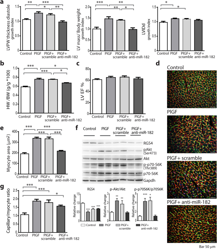
Figure 3. Inhibition of miR-182 prevents myocardial hypertrophy and Akt/mTORC1 activation.
(a) Growth index of echo-determined: LV posterior wall (LVPW) thickness, LV mass (normalized to body weight) and LV internal diameter in diastole (LVIDd) in untreated PlGF mice and PlGF mice treated with either anti-miR-182 or miR-scramble, compared with control. (b) Heart weight (HW)/body weight (BW) ratio at the end of treatment. (c) Echo-determined LV ejection fraction (LVEF). (d) Representative LV myocardium sections immunostained with anti CD31 and anti-laminin Abs. (e) Cardiomyocyte cross-sectional area measurements. (f) Western blot analysis of RGS4, AktSer473, p70-S6KThr389 expression in LV tissue lysates. (g) Capillary/myocyte ratio. n = 9 (controls); 9 (PlGF); 6 (PlGF+miR-scramble); 6 (PlGF+anti-miR-182). *P < 0.05; **P < 0.01; ***P < 0.001 vs. control or as indicated.

