Fig. 6.

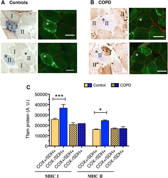
Abnormal expression pattern of TFAM protein in individual COX-deficient (COX−/SDH+) fibers of COPD locomotor muscle. a–b Representative images of muscle immunofluorescently labeled for TFAM showing higher levels of a TFAM protein in COX-deficient fibers compared to COX-normal fibers in vastus lateralis muscle of control subjects and b no difference in TFAM protein between COX-normal and COX-deficient fibers in COPD patients. Slow-twitch and fast-twitch muscle fibers identified as I and II, respectively, representing myosin heavy chain (MHC) I and MHC II fibers; scale bar is 50 μm. c Quantification of TFAM protein content in individual COX-normal (COX+/SDH+) and COX-deficient (COX−/SDH+) fibers reveals higher content in COX-deficient compared to COX-normal slow-twitch (***P < 0.001) and fast-twitch fibers (*P < 0.05) within control muscle, but no such difference in COPD muscle
