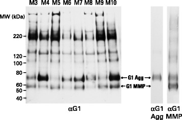
Fig. 3.

SDS-PAGE and immunoblotting of aggrecan from different individuals. Proteoglycan from guanidine extracts of OA articular cartilage, treated with keratanase and chondroitinase ABC, were analyzed by electrophoresis on polyacrylamide gels, and the fractionated proteoglycan then transferred to nitrocellulose membranes. Aggrecan was visualized by immunoblotting using an antibody recognizing the G1 region. Cartilage samples were obtained midway between the lesion and the joint margin from the femoral condyles and representative samples from eight individuals (M3-M10) are shown. Molecular weights of reference proteins are indicated at the left hand side of the blot. The migration positions of G1 generated by MMP action (G1-MMP) or aggrecanase action (G1-Agg) were determined using anti-neoepitope antibodies specific for their C-terminal peptide sequences (αG1 MMP and αG1 Agg, respectively). αG1MMP shows both terminal neoepitope and some internal sequence recognition
