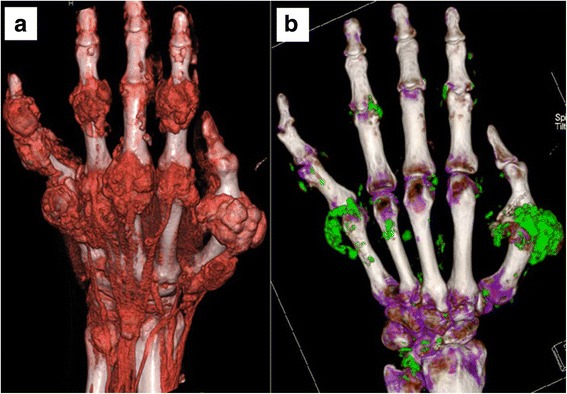
Fig. 1.

Patient 20. Large tophi are evident in the fingers with 3D volume rendering of the 2-D CT images, using proprietary software for the digital reconstruction and application of colors and varying degrees of transparency to the tissues panel (a). With DECT scanning panel (b), MSU deposits (evident by their green coding) conform to the areas of tophaceous deposits, but are sparse in some affected areas and absent in others (e.g. little finger DIP and long finger PIP)
