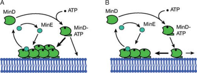
Fig. 2. Schematic representation of the Min dynamics in CA and AC models.
A. Reaction diffusion with CA. Cytoplasmic MinD (green) binds to ATP (black) and a subsequent conformational change increases its affinity for binding to the membrane. Attachment to the membrane occurs preferentially in regions already covered by MinD (thick arrow). MinE (blue) binds to membrane-bound MinD. It induces ATP hydrolysis, which eventually leads to detachment of the MinDE complex from the membrane.
B. Aggregation current. In its ATP-bound form, MinD attaches anywhere to the membrane. Attractive interactions between membrane-bound MinD moving on the membrane lead to the formation of MinD aggregates (thick arrow). MinE binds to membrane-bound MinD and releases it from the membrane as in CA models. Note that the preferential movement of membrane MinD towards MinD aggregates is the principal difference between the CA and AC models: in CA models, membrane movement either is absent or occurs purely by diffusion.

