Fig. 1.—
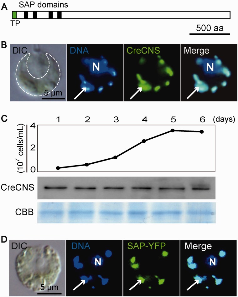
CreCNS is the constitutive cp nucleoid protein. (A) Schematic model of CreCNS. Black and green boxes represent SAP domain and transit peptides, respectively. (B) Differential interference contrast microscopy (DIC) shows a C. reinhardtii cell containing a single green cup-shaped cp (indicated by the dashed line). Indirect immunofluorescence microscopic imaging of an individual cell showing the localization of CreCNS to the cp nucleoids. Nuclear (N) and cpDNA were stained with DAPI. The CreCNS protein was detected as green signals using an anti-CreCNS primary antibody and Alexa488-conjugated secondary antibody. Arrows indicate a cp nucleoid. (C) Time-course study of cellular CreCNS accumulation analyzed by immunoblotting. Cells were cultured under continuous light. The Coomassie Brilliant Blue (CBB) stained gel is a loading control. (D) The chimeric CreCNS protein (SAP-YFP) is localized to cp nucleoids in an individual cell of C. reinhardtii. SAP-YFP was detected as green signals in indirect immunofluorescence microscopy using an anti-YFP primary antibody and Alexa488-conjugated secondary antibody.

