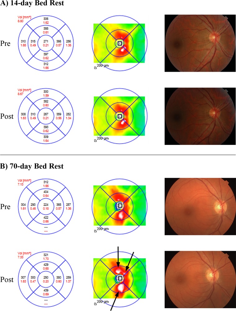
Figure 3.
Pre/post–bed rest Spectralis OCT peripapillary retinal thickness (left, black) and volume (left, red), retinal thickness map (middle), and color fundus photography (right) of two sample eyes from the 14- and the 70-day bed rest campaigns. After 14-day bed rest (A), Spectralis OCT displays minimal changes. Instead, peripapillary retinal thickening (mean increase from baseline: +18 μm [+5.3 %]) following 70-day head-down-tilt bed rest is visible on Spectralis OCT retinal thickness map as an enlargement of the white areas ([B] arrows). In both case examples, no changes are detectable on color fundus photography after bed rest.

