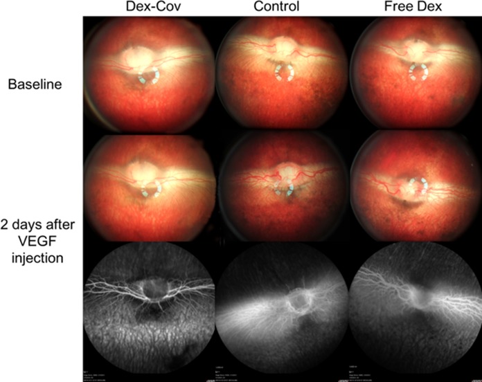
Figure 8.
Color fundus and FA images of rabbits at 4-week time point showing the effect of added VEGF on treated and untreated eyes. Vascular endothelial growth factor was administered by intravitreal injection 4 weeks after the injection of therapeutic pSiO2-COO-Dex (“Dex-Cov”), empty pSiO2 (“Control”), or free dexamethasone (“Free Dex”), as indicated. Severe vessel dilation, tortuosity, and leakage were induced by VEGF in the control and free Dex groups, whereas the pSiO2-COO-Dex group appears normal. Top (“baseline”) corresponds to treated eyes before VEGF injection (4 weeks after therapeutic injection), and the bottom two panels are images obtained 2 days after VEGF injection (4 weeks + 2 days after therapeutic injection).

