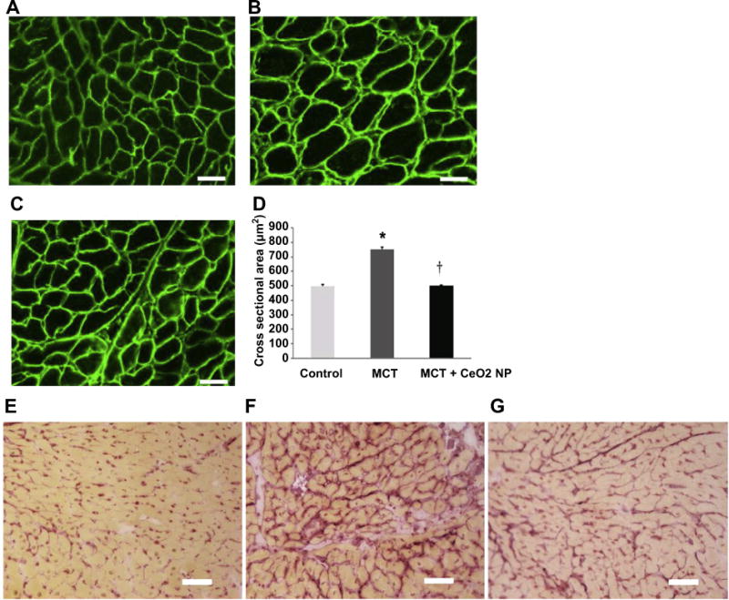
Fig. 4. CeO2 nanoparticle administration attenuates monocrotaline–induced increases in cardiomyocyte cross sectional area and cardiac fibrosis.

Dystrophin stained right ventricular sections from control (Panel A), MCT only (Panel B), and MCT + CeO2 nanoparticle treatment group (Panel C). Quantification of cardiomyocyte cross sectional area (Panel D). Picrosirius red staining was used to evaluate cardiac fibrosis in the right ventricles of control (Panel E), MCT only (Panel F) and MCT + CeO2 nanoparticle treatment groups (Panel G). Scale bar = 50 μm. Data are mean ± SEM (n = 3 rats/group). * Significantly different from control. † Significantly different from the MCT only group (P < 0.05).
