
Fig. 8. Immunoblot analysis of RV tissue demonstrates attenuation of monocrotaline induced increase in markers of oxidative stress by cerium oxide nanoparticles.
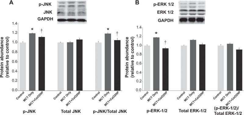
Protein isolates of RV from control, MCT only and MCT + CeO2 nanoparticles treated group was analyzed by immunoblotting to determine the amount of p-JNK, total JNK(Panel A), p-ERk1/2, and total ERK1/2 (Panel B) normalized to the expression of GAPDH. * Significantly different from control. † Significantly different from the MCT only group (P < 0.05). n = 6 animals/group.