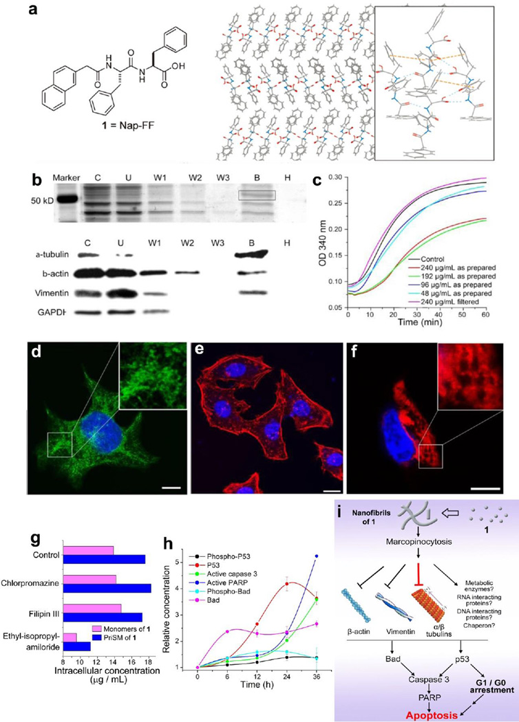
Figure 3.
(a) The chemical structure of 1 and a possible molecular arrangement in the nanofibers of 1 based on its crystal structure. (b) Molecular hydrogel protein binding (MHPB) assay. Upper panel: Silver stain of SDS-PAGE shows a major protein band at ~ 55 kDa in lane B. lower panel: Western blot indicates cytoskeletal proteins as the primary molecular targets. (c) Tubulin polymerization assays with 1. (d, e, f) Confocal images show the assemblies of 1 impede the dynamic of cytoskeletal proteins. (g) Cellular uptake of 1 in HeLa cells treated by endocytosis inhibitors. (h) Time dependent activation of apoptotic proteins of HeLa cells treated with 1. (i) The mechanism of the selective cytotoxicity of 1 towards cancer cell.[38, 44] (Print with the permission from © 2013 Wiley-VCH Verlag GmbH & Co. KGaA, Weinheim)

