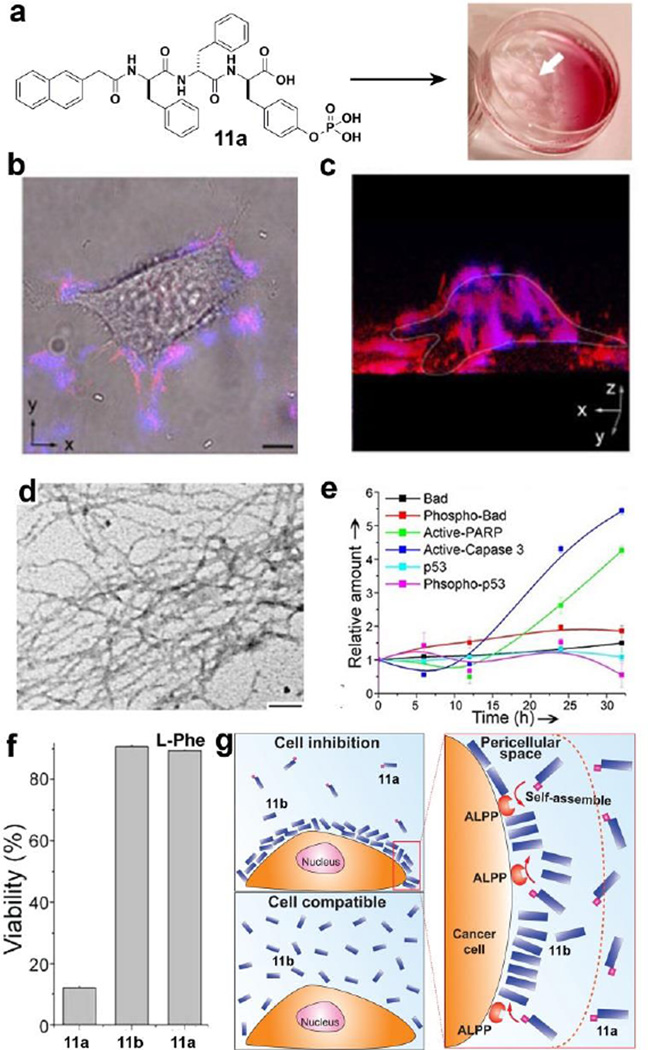
Figure 9.
(a) HeLa cells incubated with precursor 11a (560 µM) for 2h afford a hydrogel on the cells. (b) Overlaid images and (c) 3D stacked z-scan images of Congo red and DAPI stained HeLa cells after the incubation of the HeLa cells with 11a for 12h. (d) TEM images of the pericellular nanofibers on the HeLa cells treated by 11a (280 µM). (e) Change of relative amount of apoptosis signal molecules over time in HeLa cells treated by 11a (280 µM). (f) Cell viability of HeLa cells incubated with 11a (280 µM), 11b (280 µM), and 11a (280 µM) plus L-Phe (1.0 mM). (g) Enzyme-instructed self-assembly to form pericellular nanofibers/hydrogel and selectively induce cell death.[56, 57] (Print with the permission from © 2004 Wiley-VCH Verlag GmbH & Co. KGaA, Weinheim)

