Figure 3.

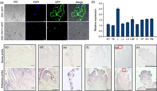
Subcellular localization and expression pattern analysis of PAY1. (a) PAY1 subcellular localization. 35S::GFP (top) and 35S::PAY1–GFP fusion gene (bottom) were transiently expressed in tobacco epidermal cells. The PAY1–GFP fusion protein was exclusively expressed in the nucleus. Scale bars, 100 μm. (b) The relative expression levels of PAY1 in various organs. RT, root; TB, tiller base; L, leaf; LJ, lamina joint; LS, leaf sheath; LSP, leaf‐sheath pulvinus; C, culm; YP, young panicle; SH, spikelet hull; PB, panicle branch. (c–h) PAY1 expression patterns revealed by mRNA in situ hybridization. The top panels are sense probes as negative controls, and the bottom panels are antisense probes. (h) was enlarged from (g) marked by red square. Scale bars, 200 μm.
