Figure 5.

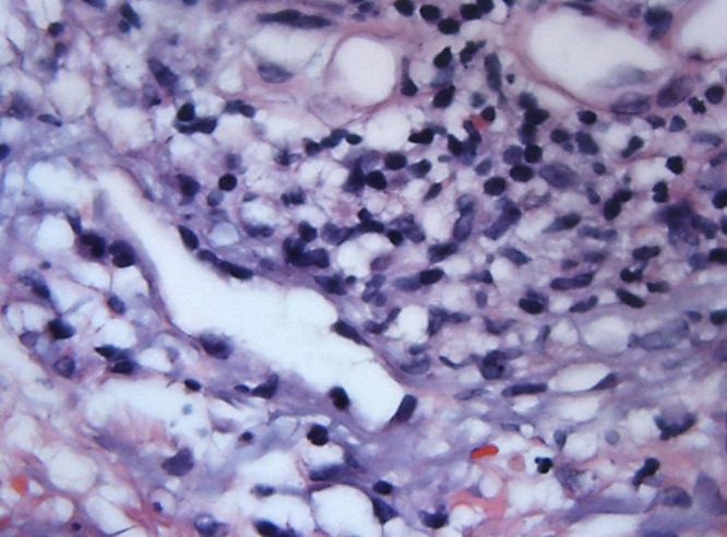
63× view demonstrating showing a perivascular and interstitial infiltrate consisting of lymphocytes, red blood cells and histiocytoid cells. [Copyright: ©2016 Shalaby et al.]

63× view demonstrating showing a perivascular and interstitial infiltrate consisting of lymphocytes, red blood cells and histiocytoid cells. [Copyright: ©2016 Shalaby et al.]