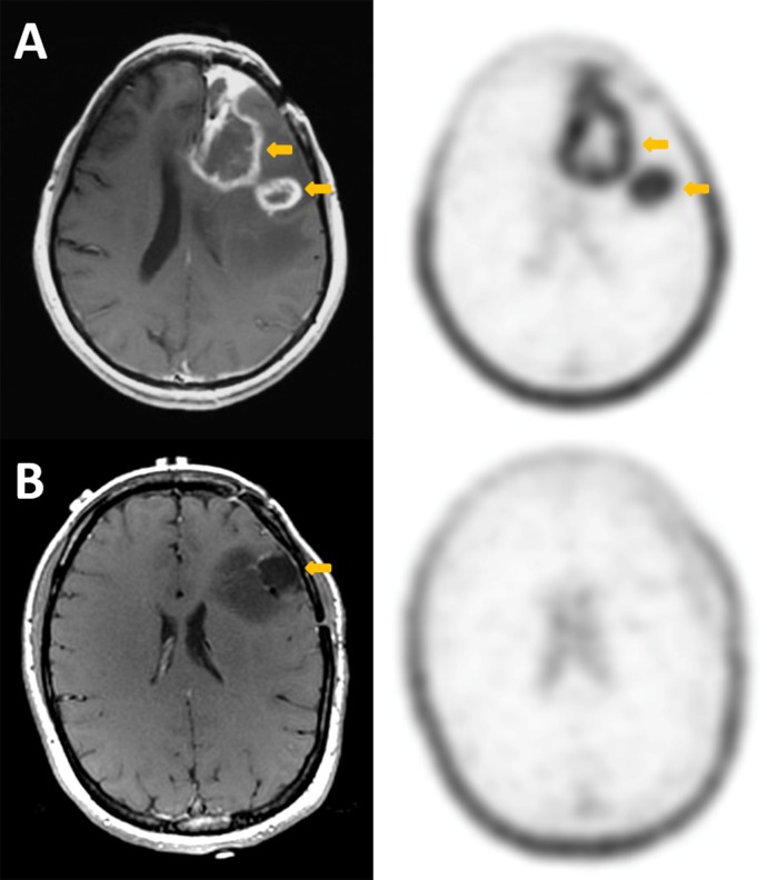
Fig 4. Comparison of axial post-contrast T1 MRI (left column) and 18F-FSPG PET (right column) in the two subjects with primary brain tumor.
Subject A has recurrent high-grade glioblastoma, while subject B has an enlarging, partially resected low-grade oligodendroglioma. In each case, the arrows point to the primary lesions. For subject A, there is intense accumulation of 18F-FSPG in the enhancing lesion (SUV 5.2), while in subject B, there is no accumulation of the radiotracer (SUV 0.7) in the non-enhancing lesion. Comparatively, the background brain SUV is 0.1 for both subjects. There is incidental note of prominent physiologic uptake of 18F-FSPG in the scalp.

