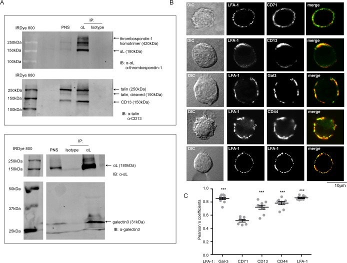
Fig 3. Validation of MS results by WB and proximity study by confocal microscopy for selected proteins.
(A) Protein complexes of LFA-1, thrombospondin-1, talin-1, CD13 (top) and galectin-3 (bottom) in imDCs (day6) were co-immunoprecipitated with LFA-1. LFA-1 was enriched using mAb (clone SPV-L7) directed against αL (CD11a). mIgG1 coated beads were included as control IP. PNS: post nuclear supernatant. Samples were analysed in non-reducing conditions. (B) Confocal microscopy analysis of co-capping of LFA-1 and galectin-3, CD44 and CD71 on imDCs (day6). Receptor co-capping and staining were performed as described in Material and Methods. Antibodies against LFA-1 (clones: NKI-L15 and TS2/4) and CD71 are positive and negative markers for co-localization, respectively. Results are representatives of multiple cells per condition (n>10.) in two independent experiments. (C) To quantify the degree of co-localization between LFA-1 and binding candidates, Pearson´s coefficient was calculated. The values can vary between 0 and 1 (1 = 100% colocalization). P-values were compared to co-capping of LFA-1 with CD71 by two-tailed t-test, *** <0.001. Co-capping and staining were performed as described in Materials and Methods.

