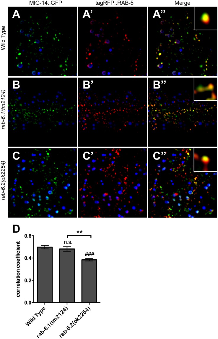
Fig 10. RAB-6.1 and RAB-6.2 drive MIG-14 out of early endosomes.
Fluorescent images were acquired in (A-A”) wild-type animals, (B-B”) rab-6.1(tm2124) mutants, or (C-C”) rab-6.2(ok2254) mutants expressing (A,B,C) MIG-14::GFP and (A’,B’,C’) tagRFP::RAB-5. Intestinal autofluorescent lysosome-like organelles are shown in the DAPI channel (blue). MIG-14::GFP colocalized with tagRFP::RAB-5 labeled early endosomes in wild-type animals (A-A” and enlarged inset), rab-6.1(tm2124) mutants (B-B” and enlarged inset), and rab-6.2(ok2254) mutants (C-C” and enlarged inset). (D) Quantification of tagRFP::RAB-5 and MIG-14::GFP colocalization as measured through average correlation coefficient. Bar, 5 μm. Error bars are SEM. N = 20. ANOVA with Dunnett’s multiple comparison to wild type (###p<0.001) or Bonferroni Multiple Comparison test (**p<0.01).

