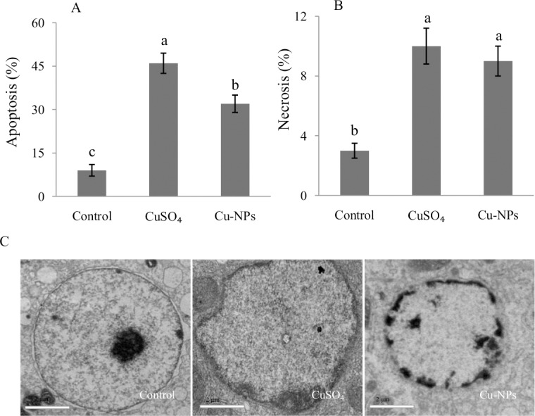
Fig 4. Cell apoptosis and necrosis in the primary hepatocytes of juvenile E.coioides after CuSO4 or Cu-NPs exposure.

(A) cell apoptosis; (B) cell necrosis; (C) the nuclear ultrastructure examined by transmission electron microscopy (TEM), scale bar = 2 μm. Data are means ±SD (n = 3). Significant differences (p <0.05) among treatments were indicated by different letters.
