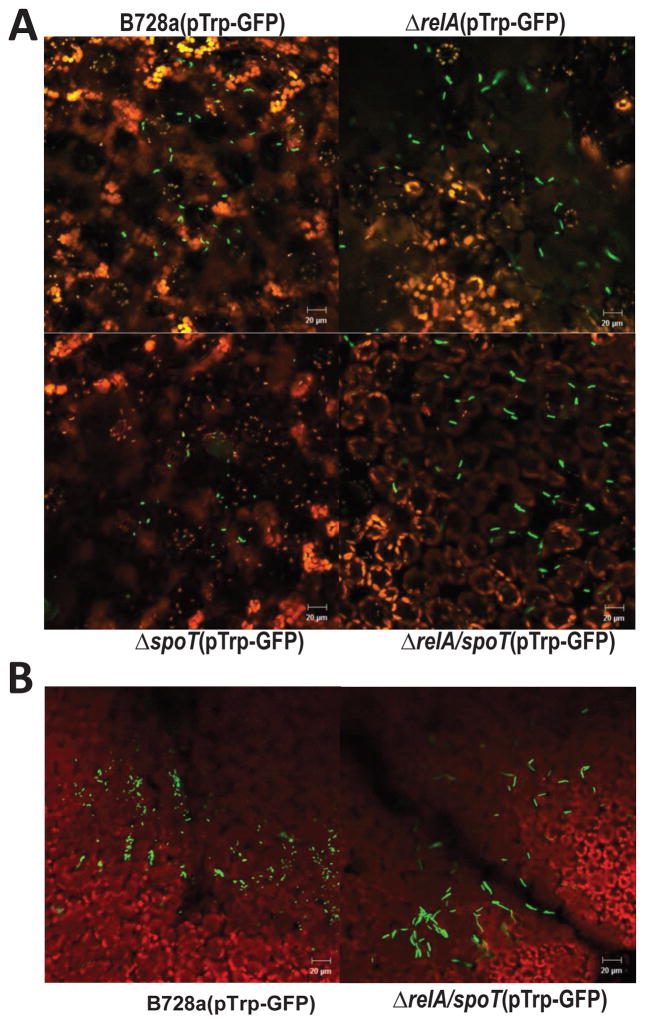
Fig. 7. ppGpp controls cell size survival on plants.
(A)Microscopy images of P. syringae pv. syringae B728a (PssB728a), relA, spoT, and relA/spoT mutant strains constitutively expressing green fluorescent protein (GFP) observed by confocal laser scanning microscopy 3 h following spray inoculation on bean leaf surfaces; (B) Microscopy images of PssB728a and relA/spoT mutant strain constitutively expressing GFP observed by confocal laser scanning microscopy 24 h post spray inoculation on bean leaf surface. GFP-labeled bacterial cells are green, and leaf cells are either red or black in color. Pictures were taken at 200x magnification with a scale bar of 20 μm. The experiments were repeated three times with similar results.

