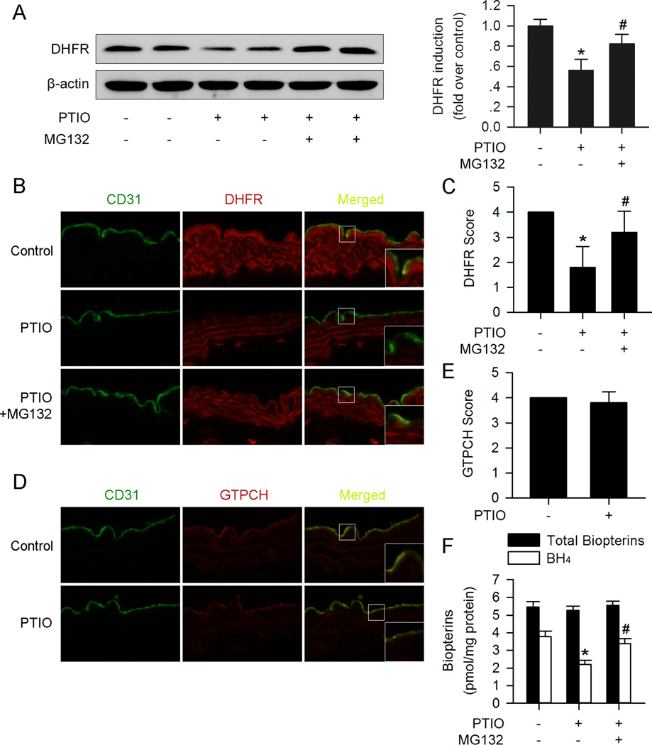
Figure 5. PTIO reduces aortic endothelial DHFR expression and BH4 content via proteasomal degradation ex vivo.
(A) Western blot analysis showed reduced DHFR expression in PTIO- (150µM) treated aortas, which could be blocked by MG132 (1µM, 6h) supplementation. (B) Representative immunofluorescence staining of DHFR (red) and endothelium marker CD31 (green) of ex vivo cultured aortas. (C) PTIO (150µM) reduced endothelial DHFR expression, while MG132 (1µM, 6h) reversed the effect. (D) Representative immunofluorescence staining of GTPCH (red) and endothelium marker CD31 (green) of ex vivo cultured aortas. (E) PTIO (150µM) had no significant effect on endothelial GTPCH expression. (F) PTIO (150µM) reduced BH4 content, which could be reversed by addition of MG132 (1µM, 6h) in aortas ex vivo. (n=4 for each group; *p<0.05 vs. control; #p<0.05 vs. PTIO)

