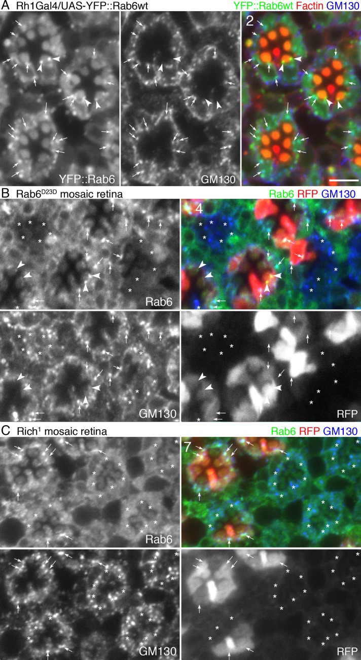
Fig 6. Rab6 localizes on Golgi and post-Golgi vesicles.
(A) Immunostaining of wild type eyes expressing YFP::Rab6wt driven by Rh1-Gal4 stained with anti-GM130 (blue) and phalloidin (red). YFP::Rab6wt is shown in green. Significant autofluorescence was also observed in pigment granules in the green channel. (B) Rab6D23D mutant mosaic eye immunostained by anti-Rab6 (GP1) (green) and anti-GM130 antibodies (blue). RFP (red) indicates wild type cells. Asterisks show Rab6D23D mutant photoreceptors. (C) Rich1 mutant mosaic eye immunostained by anti-Rab6 (GP1) (green) and anti-GM130 antibodies (blue). RFP (red) indicates wild type cells. Asterisks show Rich1 mutant photoreceptors. Scale bars: 5 μm (A–C). Numbers of the samples observed were shown in the top-left corner of the composite images.

