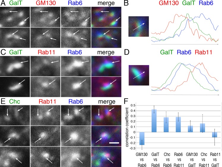
Fig 7. Rab6 localizes at the trans-cisternae/trans-Golgi network and Rab11-positive compartment.
(A) Immunostaining of wild type eye expressing CFP-GalT (green) driven by GMR-Gal4 stained with anti-GM130 (red) and anti-Rab6 (GP1) (blue). (B) Intensity plots of signal intensity (y-axis) versus distance in mm (x-axis) show the occurrence of overlap between channels. Respective signals from the cis-Golgi markers GM130 (red), trans-Golgi marker CFP-GalT (green) and Rab6 (blue). (C) Immunostaining of wild type eye expressing CFP-GalT (green) driven by GMR-Gal4 stained with anti-Rab11 (red) and anti-Rab6 (GP1) (blue). (D) Intensity plots of signal intensity (y-axis) versus distance in mm (x-axis) show the occurrence of overlap between channels. Respective signals from the CFP-GalT (green), Rab6 (blue), and recycling endosome marker Rab11 (red). (E) Immunostaining of wild type eye by anti-Chc (green), anti-Rab11 (red), and anti-Rab6 (GP1) (blue). (F) Pearson’s correlation coefficients of the co-localization of the indicated protein sets. Scale bars: 1 μm (A, C, E).

