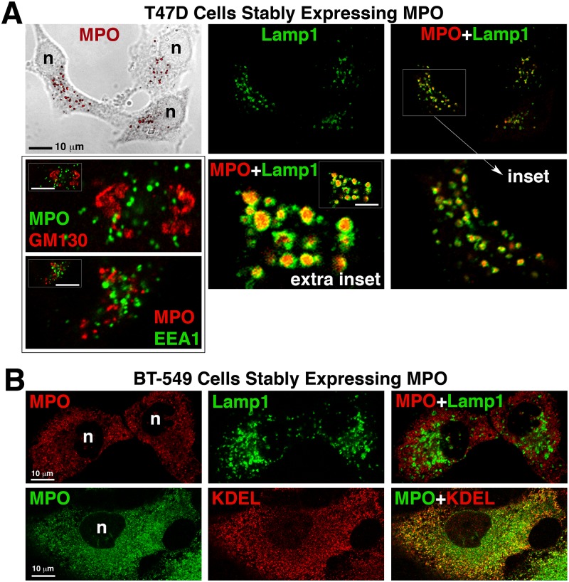
Fig 4. Subcellular localization of MPO in cells that DO (T47D) and DO NOT (BT549) produce heterotetramer.
Cells grown on uncoated glass coverslips were fixed, permeabolized and double-labeled with antibodies against the indicated proteins. Mounted coverslips were imaged with a 63x oil objective on a Zeiss LSM 710 confocal microscope. (A) Fluorescent images of the T47D-MPO stable cell line showing that MPO colocalizes with the lysosomal membrane protein Lamp1, but not with the early endosome protein EEA1 or the cis-Golgi protein GM130. An extra inset of a cell with very large lysosomes is included to more easily visualize that MPO-immunoreactivity (red) is concentrated within the lumen of the lysosomes. (B) Fluorescent images of the BT549-MPO stable cell line showing that MPO does not colocalize with the lysosome marker Lamp1 in these cells, but, the punctate staining pattern does partially co-localize with the endoplasmic reticulum marker KDEL. (n) nucleus.

