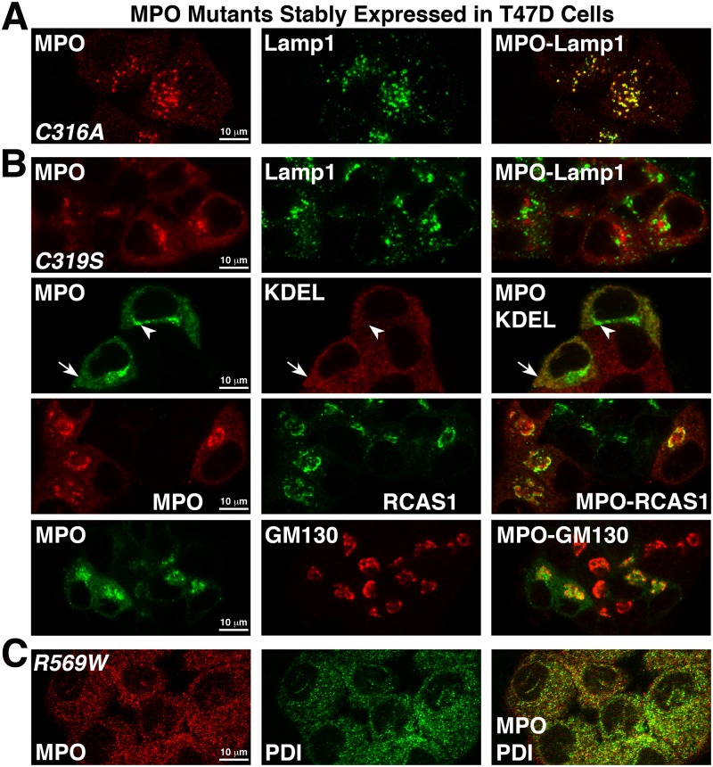
Fig 8. Subcellular localization of the C316A, C319S and R569W MPO mutants stably expressed in T47D cells.
Cells grown on glass coverslips were fixed, permeabilized and double-labeled with polyclonal MPO antibodies and the indicated organelle marker antibody. Mounted coverslips were imaged with a 63X objective on a Zeiss LSM 710 confocal microscope. (A) T47D-C316A cells labeled with antibodies against MPO and Lamp1 and shows that the C316A mutant accumulates normally within lysosomes. (B) T47D-C319S cells labeled with antibodies against MPO (all panels) and Lamp1 (top panel), the ER marker KDEL (second panel), the trans-Golgi marker RCAS1 (third panel) and the cis-Golgi marker GM130 (fourth panel). Distinct localizations of the C319S mutant to both the ER (arrow) and Golgi (arrowhead) are indicated on the second panel. (C) T47D-R569W cells labeled with antibodies against MPO and the ER marker protein disulfide isomerase (PDI).

