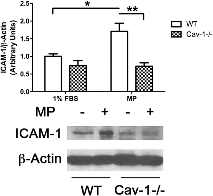
Fig 2. MP-induced ICAM-1 expression requires caveolin-1/caveolae.
WT and Cav1-/- MLEC’s were treated with MPs (Approx 40,000 MPs/mL) for 24 hrs. Cells were collected, homogenized, lysed and total cellular protein separated by 5–15% SDS-PAGE followed by Western blotting to detect ICAM-1 and β-Actin. Densitometric quantification showed ~2 fold increase in ICAM-1 expression in WT cells in response to MPs. In Cav-1-/- cells, basal expression of ICAM-1 was lower than that detected in WT cells and it remained unchanged in response to MPs. (Avg ± SEM two-way ANOVA and bonferroni’s post hoc analysis, n = 8, *p<0.05, **p<0.01).

