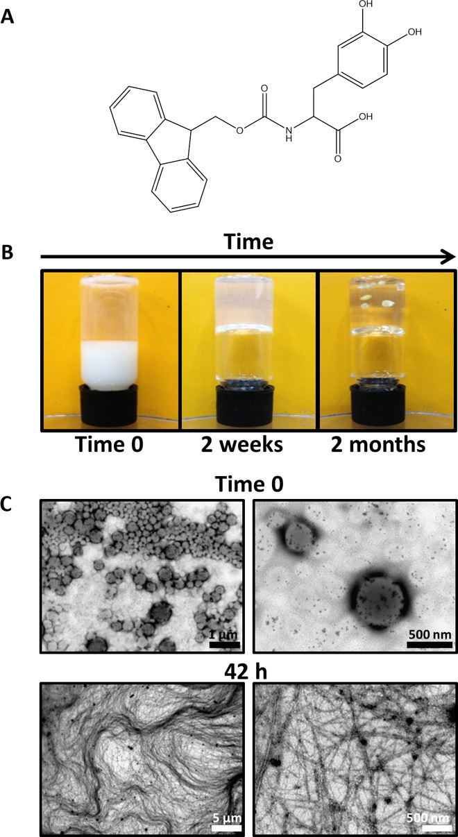
Fig. 1. Macroscopic view and imaging of Fmoc-DOPA assembly process.

(A) Chemical structure of Fmoc-DOPA. (B) Three macroscopically observable changes of Fmoc-DOPA preparation over time: turbidity change, gaining gel-like viscoelastic characteristics as indicated by a simple qualitative vial inversion assay, and spontaneous formation of crystals in the gel (from left to right). (C) Transmission electron microscopy (TEM) micrographs of Fmoc-DOPA samples taken immediately upon the initiation of assembly (time 0) and 42 hours after the initiation of the assembly at the semitransparent gel-like state.
