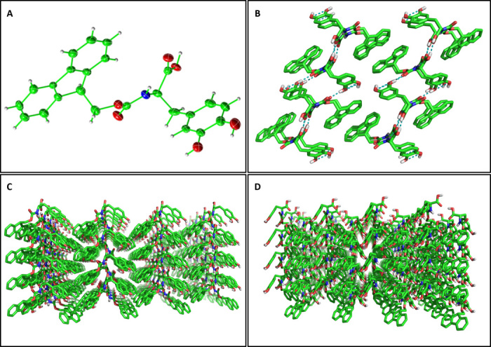
Fig. 5. X-ray structure of Fmoc-DOPA single crystals.

(A) View of the asymmetric unit, showing a single Fmoc-DOPA molecule with thermal ellipsoids at 50% probability level. (B) Crystal packing of 16 molecules along the crystallographic b axis. The interlocking hydrogen bonding network is shown in teal. (C) Extended crystal packing along the crystallographic a axis. (D) Extended crystal packing along the crystallographic c axis.
