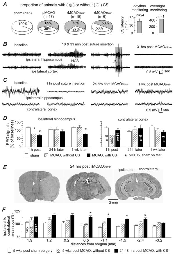
Fig. 4. Early-onset CS and related measures in adult mice.
Data were collected from adult mice (6–8 months-old). A, CS incidences in different procedural groups (left) and CS latencies (right). Only one adult mouse had CS onset during overnight video monitoring. B, representative EEG traces collected from one animal via local differential recordings from the ipsilateral hippocampus and parietal cortex). Left to right, signals collected during baseline monitoring, 10 min post surgery, 31 min post surgery, and 3 hours following rMCAO45min. C, representative EEG traces collected from another animal via monopolar recordings from the ipsilateral hippocampus and contralateral cortex. Left to right, signals collected during baseline monitoring, 1 hour post surgery, 24 hours post rMCAO90min, and 1 week later. D, the root mean square (RMS) of EEG signals measured from data segments collected during baseline monitoring and at 1 hour, 24 hours and 1 week post sham operation or post MCAO. Data were presented as a percentage of the baseline EEG signals and grouped for animals with or without CS following sham operation or pMCAO/rMCAO. E, images of cresyl violet-stained brain sections obtained from one animal 24 hours post rMCAO90min. Note the weakly stained ipsilateral regions and enlarged ipsilateral to contralateral hemispheric areas in left and middle images. F, ipsilateral and contralateral hemispheric areas measured from brain sections at 8 coronal levels (from bregma −3.2 mm to 1.9 mm). Area of the ipsilateral hemisphere was normalized as a percentage of the corresponding contralateral hemisphere, and data were grouped for sham controls and animals with or without CS following either pMCAO or rMCAO. *, sham controls vs. other groups, p<0.05, one way ANOVA.

