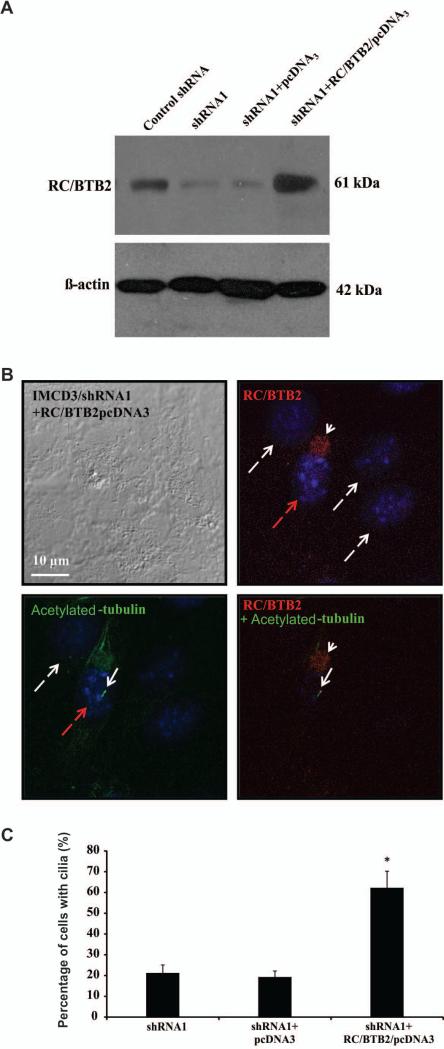
Figure 6. Re-expression of RC/BTB2 in the stable IMCD3 knockdown cells rescue ciliogenesis defect phenotype observed in the stable knockdown cells.
A. Western blot analysis of RC/BTB2 expression in the transfected stable shRNA knockdown IMCD3 cells. Notice that endogenous RC/BTB2 expression was dramatically reduced in the stable shRNA1 cells. However, its expression was increased when these cells were transfected with RC/BTB2/pcDNA3 plasmid, but not the empty pcDNA3 plasmid.
B. Representative images of the RC/BTB2 stable knockdown cells transfected with RC/BTB2/pcDNA3 plasmid stained by an anti-RC/BTB2 antibody and an anti-acetylated tubulin antibody. RC/BTB2 was labeled in red (white arrow head) and acetylated tubulin in green (white arrow). Notice that the cell expressing RC/BTB2 (with red dashed arrow) forms a normal like cilium. No cilia were observed in the cells without RC/BTB2 signal (white dashed arrows).
C. Percentage of cells with cilia with or without RC/BTB2 expression in the stable knockdown IMCD3 cells (shRNA1) transfected with RC/BTB2/pcDNA3 plasmid or empty pcDNA3 plasmid.
In each experiment, the cell number with cilia was counted from 100 cells with RC/BTB2 expression and 100 without RC/BTB2 expression in the RC/BTB2/pcDNA3 transfected cells, and 100 cells transfected with empty pcDNA3 plasmid, and percentages were calculated. The data were from three independent experiments. *p<0.05.

