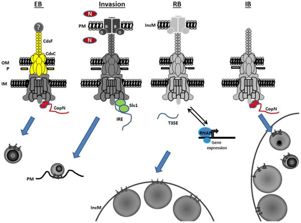
Figure 1.
Working model for T3S during chlamydial development. A completely assembled T3SA is present in the EB and spans the inner membrane (IM), periplasmic P-layer (P), and outer membrane (OM). Secretion activity is prevented by disulfide bonding within CdsF and CdsC (shown in yellow), and by positioning of the CopN plug on the cytoplasmic face of the T3SA. Secretion activity is activated upon contact of an EB with a host cell plasma membrane (PM). This results in displacement of a potential tip protein (?), secretion of CopN, and deployment of the translocon proteins CopB and CopD. The chaperone Slc1 can then mediate prioritized secretion of invasion related effectors (IRE) that orchestrate the invasion process. T3S by intracellular chlamydiae is maintained by association of the RB with the inclusion membrane (IncM) and subsequence de novo expression of T3SA genes (light gray). Chaperone-independent secretion becomes dominant and components of the T3SA affect chlamydial gene expression through interactions with RNAP. T3S activity ceases when CopN re-associates with the apparatus concomitant with RB conversion to IncM-dissociated intermediate bodies (IM).

