Figure 2.
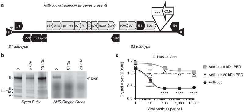
PEGylation of E3-intact Ad6 expressing luciferase. (a) Ad6-Luc vector genome structure. A luciferase expression cassette was inserted into the region between fiber and E4 sequence of Ad6. (b) Unmodified and PEGylated Ad6-Luc were separated on SDS-PAGE, virion hexon was stained with Sypro Ruby (left) and NHS-Oregon Green (right). (c) In vitro testing the viability of human DU145 prostate carcinoma cells with Ad6-Luc, Ad6-Luc 5 kDa-PEG, and Ad6-Luc 20 kDa-PEG. Cells were seeded on 96-well plates, and infected after 24 hours at triplicated multiplicities of infection (MOI) of viral particles/cell. At day 4 after infection, cells were stained with crystal violet protocol and read the absorbance at 580 nm. Statistical significance was analyzed by one-way analysis of variance. Ad6 Luc was significantly more lethal than Ad6 Luc with 5 kDa PEG at MOIs of 10 vp/cell above and was more effective than the 20 kDa PEGylated vector at MOIs of 100 vp/cell and above (P = 0.0001). At 10 vp/cell, Ad6 20 kDa PEG vector was more lethal than Ad6 5 kDa vector (P = 0.01).

