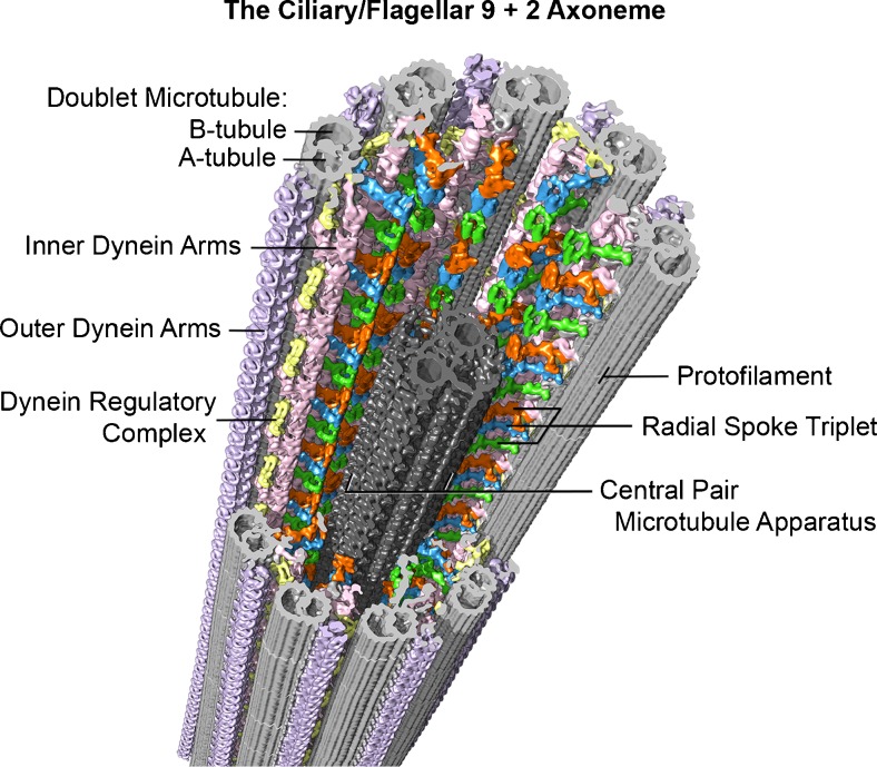
Fig. 3.
Example of the current, advanced imaging of the 9 + 2 axoneme (from sea urchin, Strongylocentrotus purpuratus, sperm flagella), using cryo-electron tomography with a resolution of approximately 3 nm. In this method, isolated flagella or axonemes, applied to special EM grids, are frozen within a few milliseconds in liquid ethane, which prevents damaging ice crystal formation. The specimen is then transferred to a cryo-transfer holder cooled with liquid nitrogen and inserted into the transmission electron microscope. After locating a promising area of a frozen flagellum or axoneme at medium magnification, a tilt series with up to 100 tilted views (from −65° to +65°) is recorded at higher magnification with low electron doses to minimize specimen radiation damage. The tilt series are then computationally aligned and the 3D structure of the specimen is reconstructed. The 96-nm longitudinal repeats of the axoneme (see text) are then extracted and averaged to increase the signal to noise ratio and thus resolution. Finally, the averaged repeat is visualized in 3D using isosurface rendering, as shown here. Some of the major structural features are labeled: Doublet A- and B-tubules (gray), radial spokes 1–3 (green, blue, orange), outer dynein arms (lavender), inner dynein arms (pink), nexin-dynein regulatory complex (yellow), and the central pair microtubule apparatus (charcoal). Image courtesy of Daniel Stoddard and Dr. Jianfeng Lin from the laboratory of Dr. Daniela Nicastro (Brandeis University and University of Texas Southwestern Medical Center). See references [10–13]

