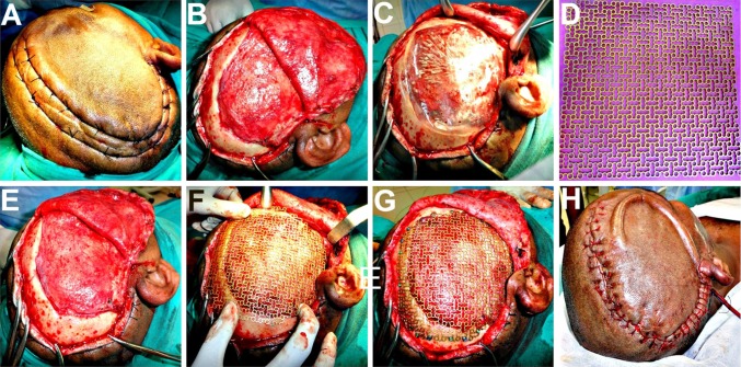
Fig. 3.
A Haemostatic sutures placed on either side of the proposed incision line, to reduce the intraoperative bleeding. B Full thickness scalp flap raised in a sub-pericranial plane around the defect and along the avascular subgaleal plane over the defect. C A prefabricated acrylic plate kept as a standby, was tried for fit. D–F Titanium mesh implant moulded to attain the appropriate curvature of the skull and adapted over the defect. G Implant cut into shape, contoured and fixed using titanium micro-screws. H A vacuum assisted closed suction drain placed prior to scalp flap closure in layers

