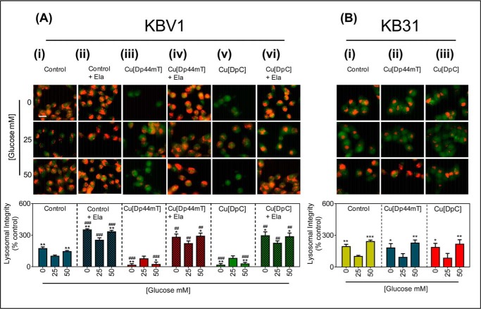
FIGURE 8.
Glucose variation-induced stress potentiates LMP by Cu[Dp44mT] and Cu[DpC] via lysosomal-Pgp. A, KBV1 (+Pgp) cells were preincubated for 2 h at 37 °C with low (0 mm), normal (25 mm), or high (50 mm) glucose media and also during the final 30 min/37 °C treatment with media with 0, 25, or 50 mm glucose alone (i), Ela (0.2 μm) (ii), Cu[Dp44mT] (20 μm) (iii), Cu[Dp44mT] (20 μm) and Ela (0.2 μm) (iv), Cu[DpC] (10 μm) (v), or Cu[DpC] (10 μm) and Ela (0.2 μm) (vi). Cells were then stained with AO (5 μm, 12 min/37 °C). B, KB31 (−Pgp) cells were preincubated under the glucose conditions as in A and then incubated for the final 30 min/37 °C treatment with media with 0, 25, or 50 mm glucose alone(i), Cu[Dp44mT] (20 μm) (ii), or Cu[DpC] (10 μm) (iii). Cells were then stained with AO as in A. Lysosomal integrity was quantified as AO (red) fluorescence intensity (as a percentage of control (25 mm) glucose) using ImageJ software. Data are the mean ± S.D. of four independent experiments. *, p < 0.05; **, p < 0.01, and ***, p < 0.001 are relative to the respective normal (25 mm) glucose (e.g. KBV1 control 0 mm glucose versus KBV1 control 25 mm glucose). ##, p < 0.01, and ###, p < 0.001 are relative to respective control glucose treatments (e.g. KBV1 control + Ela 0 mm glucose versus KBV1 control 0 mm glucose). Scale bar = 10 μm.

