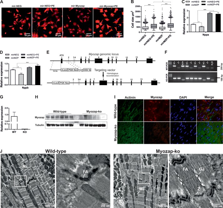
FIGURE 1.
Knockdown of myozap in NRVCMs and basal characterization of Mzp−/− mice. A, primary NRVCMs were infected with adenovirus encoding synthetic microRNA to knock down myozap in the absence or presence of PE and immunostained against α-actinin. B, cell surface area was measured using Keyence's HybridCellCount software module in fluorescence intensity single-extraction mode. Statistical significance was determined using Shapiro-Wilk (for equal distribution) and Kruskal-Wallis (one-way ANOVA) tests. n > 500. Expression of fetal genes Nppa (C) and Nppb (D) was determined by quantitative real time PCR. Data represented are mean of two independent experiments performed in triplicate. E, strategic diagram showing the targeting vector and myozap genomic locus. Homologous recombination replaces exon 1 encoding the translation initiation site with a neomycin-lacZ reporter cassette. Deletion of myozap was confirmed by genotyping (F), qRT-PCR (G), immunoblotting (H), and immunofluorescence microscopy (I). J, electron micrographs showing the structural architecture of cardiac muscle. As to the intercalated discs, fasciae adherents (FA) are shown at higher magnification (see boxes). There are no ultrastructural differences between wild-type (WT) and myozap−/− (Mzp−/−) mice. GJ, gap junction; M, M-line; Mi, mitochondria; Z, Z-line. Statistical significance was calculated by two-way ANOVA. Error bars show mean ± S.E. *, p < 0.05; ***, p < 0.001.

