FIGURE 4.
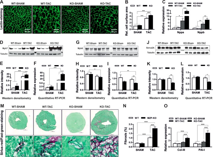
Biomechanical stress accelerated cellular hypertrophy in myozap-deficient mice. Heart slices obtained from SHAM or TAC-operated wild-type (WT) or myozap-KO (Mzp−/−) mice were stained with FITC-conjugated lectin (A), and cell surface area of the cardiomyocytes was measured (B) showing accelerated hypertrophy in Mzp−/− mice compared with the WT littermates. This was further confirmed by super induction of fetal genes Nppa/Nppb determined by quantitative real time PCR (C), marked up-regulation of β-myosin heavy chain (Myh7, D–F), and reduced expression of alpha-myosin heavy chain (Myh6, G–I), or Serca2a (J–L), both at transcript and protein levels. M, heart sections obtained from SHAM or TAC operated wild-type (WT) or Mzp−/− mice were stained with Sirius-red/Fast-green (upper panel), and the representative fibrotic area is highlighted in the lower panel. N, fibrotic lesions stained in red were measured using Keyence's HybridCellCount software module in bright field single-extraction mode, which confirms the increased fibrosis in Mzp−/− mice in comparison with WT littermates. O, increased expression of fibrotic markers collagen III (Col-III) and plasminogen activator inhibitor I (PAI-I) further supports the increased fibrosis in Mzp−/− mice. n = 6. Statistical significance was calculated by two-way ANOVA. Error bars show mean ± S.E. *, p < 0.05; **, p < 0.01; ***, p < 0.001.

