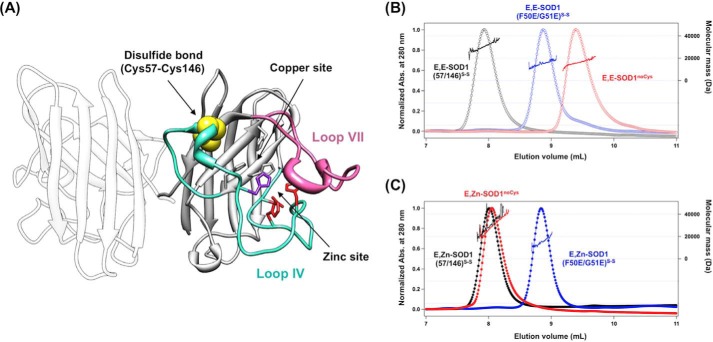
FIGURE 1.
The most immature state of SOD1 is monomeric, but its conformation is significantly different from that of an SOD1 protomer. A, a crystal structure of SOD1 homodimer (PDB ID 1HL5). Loops IV (cyan) and VII (pink) of SOD1 are indicated in the crystal structure, and the disulfide bond between Cys-57 and Cys-146 is shown in yellow. Ligands for binding copper and zinc ions are also shown: gray, His-46, His-48, and His-120 for copper binding; red, His-71, His-80, Asp-83 for zinc binding; purple, His63 for copper and zinc binding. B and C, gel filtration chromatograms of E,E-SOD1noCys (red open circles), E,E-SOD1(57/146)S-S (black open circles), and E,E-SOD1(F50E/G51E)S-S (blue open circles) (B) and E,Zn-SOD1noCys (red-filled circles), E,Zn-SOD1(57/146)S-S (black-filled circles), and E,Zn-SOD1(F50E/G51E)S-S (blue-filled circles) (C) were shown. A sample containing 60 μm protein in 100 mm Na-Pi, 100 mm NaCl at pH 7.0 was loaded on the gel filtration column, and the elution profile was monitored with absorbance (Abs.) change at 280 nm (left axis). Molecular mass obtained by MALS analysis is also shown in each chromatogram (right axis).

