FIGURE 1.
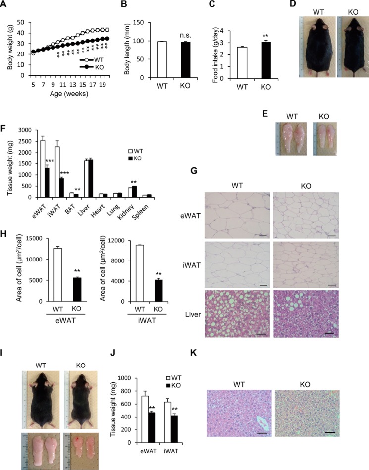
Altered fat metabolism in HFD-fed Prip-KO mice. Wild-type (WT) and Prip-KO (KO) mice were fed a HFD (A–H) or a RD (I–K) ad libitum for 16 weeks starting at 5 weeks of age. A, change in body weight of WT (n = 14) and KO (n = 14) mice. B, body length of 20-week-old WT (n = 9) and KO (n = 9) mice. C, daily food intake of 18-week-old WT (n = 7) and KO (n = 7) mice. D–H, 20-week-old WT and KO mice were used for the experiments. Representative images of overall appearance (D), epididymal WAT (E), and hematoxylin and eosin-stained sections of epididymal WAT (eWAT), inguinal WAT (iWAT), and the liver (G) are shown. Similar data were obtained from the 8 mice of each genotype (D and E). Mean tissue weights for WT (n = 8) and KO (n = 8) mice were measured (F). Mean cell sizes in eWAT (WT and KO, n = 3 sections) and iWAT (WT and KO, n = 3 sections) were calculated. Sections from 3 different mice comprised 30 cells (H). I–K, representative images of overall appearance and epididymal WAT (I), and mean epididymal WAT weights (J; WT and KO, n = 6) of RD-fed mice. Similar data were obtained from the 12 mice of each genotype (I). Representative images of hematoxylin and eosin-stained sections of the liver are shown (K). Similar images were obtained from 3 independent experiments. Scale bar, 50 μm (G and K). The data represent the mean ± S.E. **, p < 0.01; ***, p < 0.001 versus the corresponding WT value; n.s., not significant.

